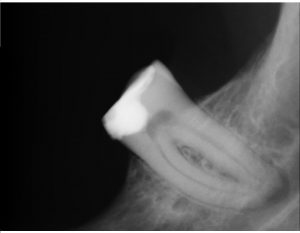
Preop view of #18. Note the PARL is not centered on the apex but extends up the distal aspect of the distal canal wall alluding to the presence of a lateral canal.
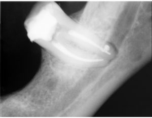
Post op image revealing the presence of a lateral canal on the distal root.
Click the link immediately below (in red) to download the endoscope video
lateral canal video
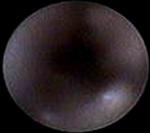
Still was taken at the 28 second mark from the above video. This image is from the apical third of the distal root (area where the lateral canal emerges). While it is subject to interpretation I would submit that the anomolous white area seen in the lower portion of the photo above is the orifice to the lateral canal seen in the post op radiograph. Agree or diagree? Shoot me an email and let me know. MLewisDMD@gmail.com


