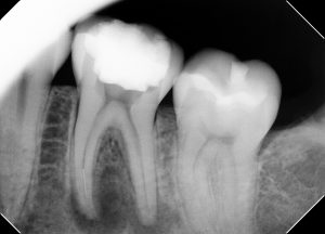
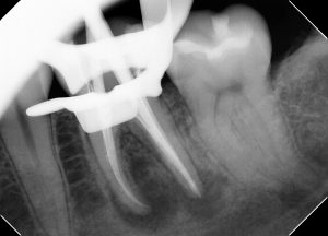
Pt referred for management of separated file in Mesial Buccal canal (forgive the cone cut)
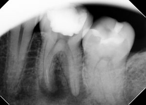
Mesailly shifted angle reveals file is located in MB canal (S.L.O.B. rule)
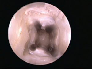
Initial appearance of chamber floor upon removal of cavit temporary. Note the narrow circular appearance fo the MB canal(upper left of photo)
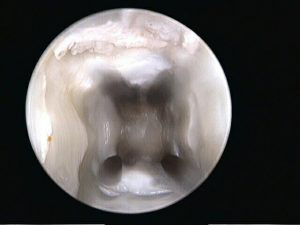
A previously missed middle mesial canal was located and debrided (compare the orifice floor to previous photo). This middle mesial canal joined up with the MB canal inthe middle third. This complex anatomy was a contributing factor for the file separating in this particular canal – file probably got got up in the narrow portion of the middle mesial canal and caused the separation. This case highlights the importance of scouting for additional canals, and establishing proper glide paths prior to instrumentation wtih rotary files.master cone image
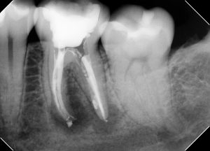
post op image after file bypass. A lateral canal fill is noted on the distal.