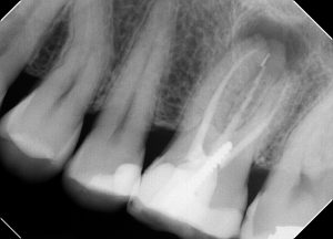
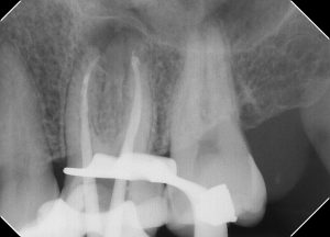
Pt presented for retreatment #14. Pre-operative image shows a separated file in the apical third of the palatal root. A large apical radiolucency is present.
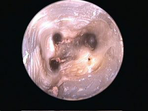
Initial access after disassembly. Note the (limited) extent of the debridement from the original RCT, specifically the circular orifice of both the MB1 canal and the palatal canal.
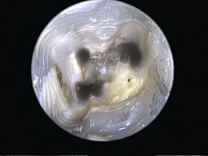
Appearance of the access cavity after proper debridement. Note the MB2 canal canal is fully opened up and the palatal canal has been broadened into it’s true elliptical form.
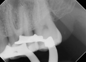
Mid op radiograph after removal of the gutta percha. The file fragment in the apical third of the palatal canal remain lodged in place.
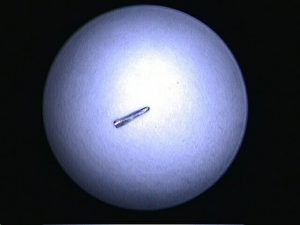
Mid op radiograph and photo showing file retrieval.
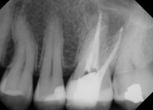
Final radiograph showing two portals of exit on the MB root and a properly cleaned and obturated palatal canal (sans file of course).

